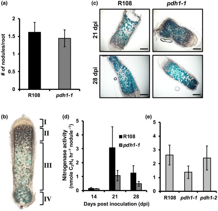
Figure 5.

The role of PDH in legume‐rhizobia symbiosis. (a) Wt and pdh1‐1 were grown on plates with low nitrogen Fahräeus medium, inoculated with S. meliloti, and the number of nodules was counted at 14 days post‐inoculation (dpi). Bars represent average number of nodules per root ± SEM., n ≥ 11. (b) Indeterminate nodules from M. truncatula consist of four developmental zones: I, meristem; II, infection; III, fixation; IV, senescence located closest to the root. These four developmental zones are shown on a characteristic nodule (enlarged for visualization) from 28 dpi. GUS staining with PnifH::UidA shows expression of bacterial nifH gene localized to the bacteroids (blue). (c) Thin sections of nodules from 21 and 28 dpi showing expression of PnifH::UidA, highlight bacteroid and nodule development in R108 and pdh1‐1. Approximately equal staining was observed in all nodule developmental zones, suggesting pdh1‐1 is not affected in nodule development or bacteroid number, scale bar = 1 mm. (d) Nitrogen fixation efficiency was measured using an acetylene reduction assay (ARA). Plants were grown under the same conditions as in (a). At 14, 21, and 28 dpi, ethylene production was measured and expressed as the average ± SEM, n > 7. (e) Nitrogen fixation efficiency assay performed in the same way as in (d), but with both mutant alleles and Wt and only at 21 dpi. Activity is expressed as the average ± SEM, n > 4
