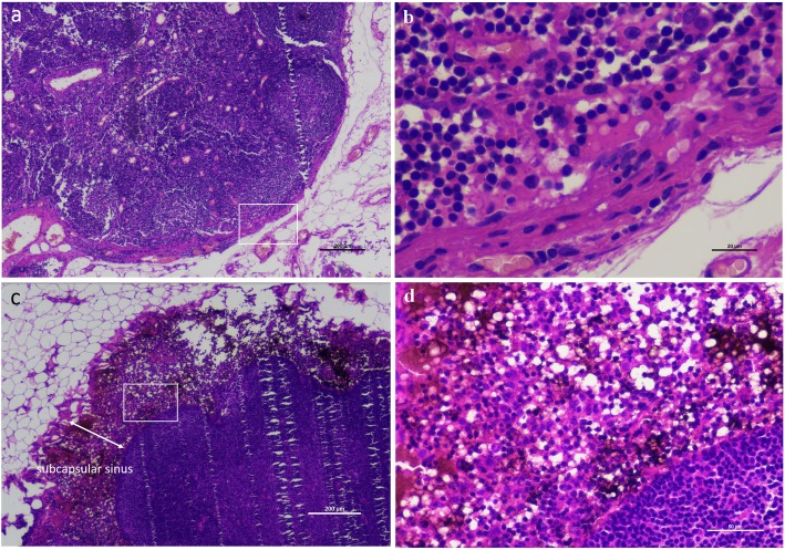
Fig. 2.
a and b Normal lymph node section without carbon nanoparticle staining. As a control sample, there was no sign of abnormal staining in the lymph node sections. c and d Lymph node section showed carbon nanoparticle staining. Histopathological examination showed that the carbon nanoparticles (the dark brown to black staining area) accumulated in the subcapsular sinus of lymph nodes in the case using carbon nanoparticles suspension. ↔indicates the subcapsular sinus area of a lymph node. Figure magnification: a × 40, b × 400, c × 40, and d × 200. b Shows the square frame area of a. d Shows the square frame area of c

