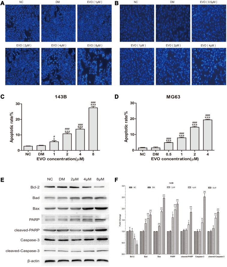
Figure 3.
EVO induces the apoptosis of 143B and MG63 cells by activating the mitochondrial apoptotic pathway. (A and B) 143B and MG63 cells were treated with different concentrations of EVO or DMSO for 24 hours, and then stained with Hoechst 33,258. (C and D) Statistical analysis for the apoptosis assay. The data are presented as the mean ± SD (n=3, each group). *p<0.05, ***p<0.001 vs NC group; #p<0.05, ###p<0.001 vs DMSO group. (E and F) Cell apoptosis‐related proteins including caspase-3, cleaved caspase-3, PARP, cleaved-PARP, Bcl-2, Bad and Bax were analyzed by Western blotting. β-Actin was served as a loading control. The data are presented as the mean ± SD (n=3, each group). *p<0.05, **p<0.01, ***p<0.001 vs NC group; #p<0.05, ##p<0.01, ###p<0.001 vs DMSO group.

