
Figure 2.
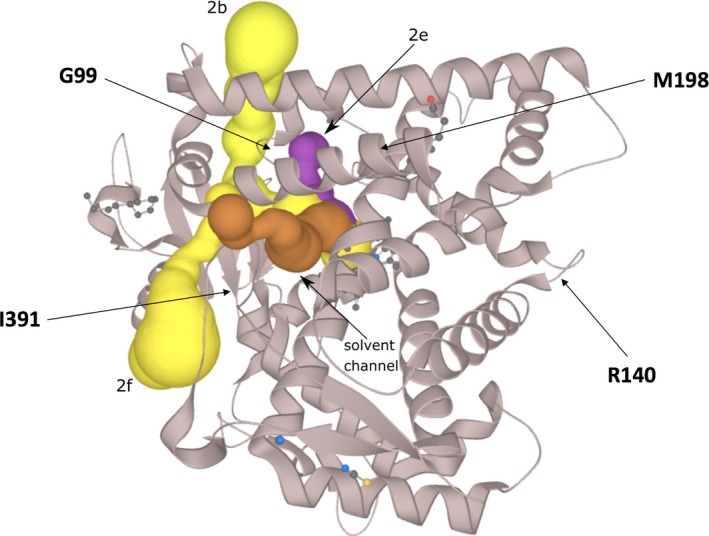
Overview of the CYP2B6 protein (PDB ID: http://www.rcsb.org/pdb/search/structidSearch.do?structureId=3IBD) with the predicted channels 2e (magenta, solid surface), 2b and 2f (yellow, solid surface). A solvent channel is shown in brown (solid surface)