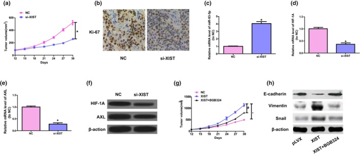
Figure 5.

XIST advances cell growth of colorectal cancer by in vivo AXL signaling. (a) The volume of tumors was determined at the range of 12 to 30 days. (b) Representative ki‐67 staining. (c‐e) The levels of HIF‐1A, miR‐93‐5p, and AXL mRNA in tumor samples. Unpaired Student's t‐test was applied to conduct statistical analysis. *p < .05 versus. NC group. (f) Protein expressions of AXL and HIF‐1A in tumor tissues were detected by western blot. (g) Subcutaneous injection was performed on mice with SW480 cells of stable transfection of XIST; the mice orally taken BGB324 twice each day. Tumor volume was detected. (h) Western blot was conducted to detect the levels of Vimentin, E‐cadherin, and Snail. One‐way ANOVA was conducted to analyze data. # p < .05 versus. XIST group, *p < .05 versus. NC group
