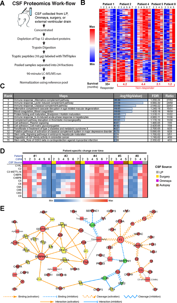
Figure 2. Proteomic analysis of serial CSF specimens from leptomeningeal melanoma metastasis patients shows protein signatures associated with poor prognosis.
A. Workflow schematic showing the overall approach for CSF preparation and analysis. B. Heatmap showing LMM-specific protein signatures that change significantly over patient treatment time and are most anti-correlated between responder and non-responders. C. Pathway enrichment analysis of data shown in B illustrates components of the complement pathway, adhesion signaling and IGF activation pathways to be enriched for in CSF of poor responders. D. A closer look at the complement signatures identified using proteomics analysis of CSF in melanoma LMM patients (from panel B). Heatmap shows the protein level changes specific to each patient’s clinical timeline. E. Experimentally consistent literature network shows the major signaling mediators upregulated in CSF of non-responders.

