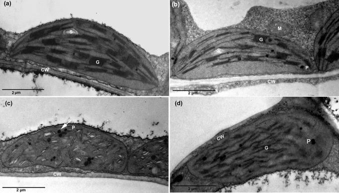
Fig. 5.
Electron micrographs of leaf chloroplast. a Distilled water treated tobacco leaf showing normal distribution of the grana (G) formed from several thylakoid membranes. b Magnetically treated distilled water tobacco leaf showing intact arrangement of the grana (G) and normal starch grain (S). c, d Tap water and magnetically treated tapwater treated tobacco leaf, respectively, showed damaged thylakoid membranes and several plastoglobules (P). Scale bars: 2 µm

