Fig. 1.
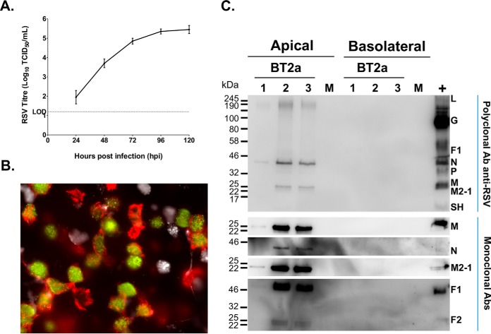
hRSV targets ciliated cells but not goblet cells, and leads to a productive infection. A, WD-PBEC cultures (n = 3 donors, in duplicate) were infected with hRSV (MOI = 0.1) or mock infected. Apical rinses were taken every 24 h and titrated using HEp-2 cells by TCID50. B, At 120 h post-infection (hpi) cultures were fixed with 4% paraformaldehyde, ciliated, goblet and infected cells were stained with anti-β-tubulin (red), anti-Muc5ac (white) and FITC-conjugated anti-RSV (green) antibodies, respectively. Cultures were examined by fluorescent microscopy at x63 magnification. C, Apical washes and basolateral medium collected at 96 hpi from infected cultures, and pooled samples from mock-infected cultures were analyzed individually by Western blotting (12 μl/lane). Ten μl/lane of a sucrose-gradient purified hRSV A2 stock was used as control. Immunoblot analyses was carried out using a polyclonal anti-hRSV antibody (upper panel) and monoclonal antibodies specific for the matrix protein (M), nucleoprotein (N), fusion (F) and M2-1 (lower panel), followed by appropriate HRP-conjugated secondary antibodies for enhanced chemiluminescence detection.

