Figure 4.
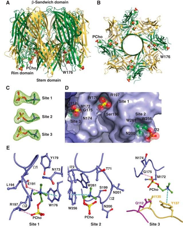
Crystal structure of C14PC in complex with the PVL heterooctamer. A and B, ribbon representation of the C14PC-bound PVL heterooctamer shown from the side (A) and cytoplasmic (B) views. The PCho moieties and the side chain of Trp176 are displayed as CPK spheres and in stick format, respectively. The LukF-PV and LukS-PV subunits are colored green and yellow, respectively. The β-sandwich, rim, and stem domains are indicated. C, 2Fo − Fc omit electron density map contoured at 1.0σ shown as green mesh around the three PCho moieties in a single protomeric unit. D, surface representation of the three PCho-binding pockets on a single protomeric unit viewed from the cytoplasmic side, with the PCho moieties shown in stick format with transparent CPK spheres. The three binding sites are labeled. The locations of key binding site residues are indicated. E, close-up view of the PCho moieties in the three binding pockets on a single protomeric unit. The side chains of residues that make direct contacts with the PCho moieties are shown in ball-and-stick format colored in blue for protomer A, in magenta for protomer G, and in yellow for protomer H. The Ω1 and Ω2 loops are labeled. Cation-π interactions are depicted as green dotted lines. Hydrogen bonds and salt bridges are represented as pink dotted lines.

