Figure 3.
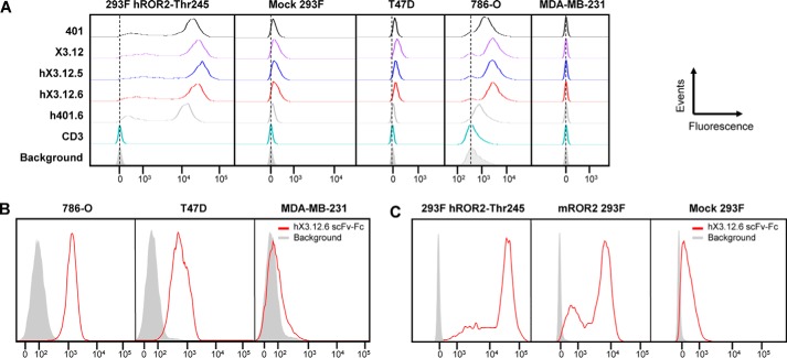
Analysis of affinity-matured and humanized mAbs by flow cytometry. A, HEK 293F cells stably transfected with human ROR2 (allotype Thr-245) were stained with 5 μg/ml of the indicated parental, affinity-matured, and humanized Fabs followed by phycoerythrin-conjugated goat anti-human F(ab′)2 pAbs. Mock-transfected HEK 293F cells served as negative control. The Fabs were also tested against T47D (ROR2+, ROR1−), 786-O (ROR2+, ROR1+), and MDA-MB-231 (ROR2−, ROR1+) cell lines. Humanized anti-human CD3 Fab v9 and secondary antibody alone (Background; gray shade) served as additional negative controls. B, after its conversion from Fab to scFv-Fc, hX3.12.6 at 5 μg/ml followed by Alexa Fluor 647–conjugated donkey anti-human F(ab′)2 pAbs was used to stain 786-O, T47D, and MDA-MB-231 cell lines. Secondary antibody alone (gray shade) served as negative control. C, flow cytometry using hX3.12.6 scFv-Fc (5 μg/ml) followed by Alexa Fluor 647–conjugated donkey anti-human F(ab′)2 pAbs for staining HEK 293F cells stably transfected with human ROR2 (allotype Thr-245) or mouse ROR2. Mock-transfected HEK 293F cells and secondary antibody alone (Background; gray shade) served as negative controls. All events were normalized to mode.

