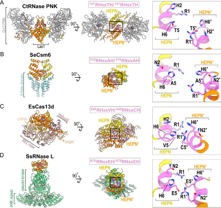
Figure 7.
Structural comparison of HEPN nucleases. A–D, ribbon diagram of (A) C. thermophilum RNase PNK (PDB ID 6OF3), (B) Staphylococcus epidermis Csm6 (PDB ID 5YJC), (C) Eubacterium siraeum Cas13d (PDB ID 6E9F) and (D) Sus scrofa RNase L (PDB ID 4O1P). HEPN-HEPN′ dimers are colored in yellow and orange along with their juxtaposed RφXXXH motifs colored in purple. Enzyme-specific insertions of RNase PNK, Csm6, Cas13d, and RNase L are colored in gray, light blue, brown, and light green, respectively. Black boxes mark the HEPN nuclease active sites formed by well-conserved HEPN nuclease motifs (purple). The inset is a zoom of the juxtaposed RφXXXH motifs forming the catalytic site. Conserved residues of the RφXXXH motifs are shown and the second copy of the RφXXXH motif is designated by prime.

