Figure 7.
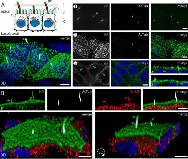
Zebrafish prom3 is distributed in a nonpolarized fashion in polarized epithelial cells and co-localized with Arl13b at the primary cilium. A and B, zebrafish prom3-HA–expressing MDCK cells growing as a polarized cell monolayer (7 dpc) were either double-immunolabeled (A) for prom3-HA using anti-HA antibody (green) and anti-AcTub antibody (white) or triple-immunolabeled (B) with the detection of Arl13b (red). Nuclei (nu) were counterstained with DAPI (blue) prior to CLSM analyses. Three to four single optical x-y section planes (0.4-μm slice each) throughout the cell monolayer as illustrated in the cartoon (A, sections 1–3) revealed the presence of prom3-HA in primary cilia (pc, sections 1 and 2, green arrow) highlighted with AcTub staining and microvilli (mv, section 2, asterisk) present at the apical membrane. Prom3-HA is also detected at lateral membranes (section 3, green arrowhead). Three-dimensional views (x-z orientation (z), top panels; top-side orientations (3D), bottom panels) were built from 42 x-y sections throughout cells. The immunolabeling revealed the co-localization of prom3-HA and Arl13b in the ciliary compartment (B, green and red arrows, respectively). Note the asymmetric distribution of prom3-HA at the ciliary membrane (B, bottom panels, green arrow). Tj, tight junction. Scale bars, 5 μm.

