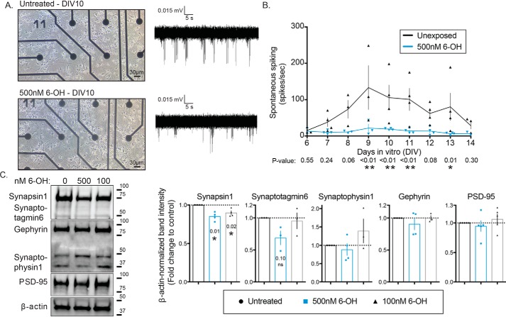
Figure 1.
Chronic 6-OH–BDE-47 exposure suppresses spontaneous neuronal activity and alters pre-synapses. Primary rat cortical neurons were grown on MEAs in order to assess the effects of 6-OH exposure on electrical activity. A, example images of neurons at DIV10 with or without exposure to 500 nm 6-OH (left), and example traces of recorded spontaneous activity (right). B, detectable activity was measured daily with 3-min recordings and quantified through the first 2 weeks of growth in vitro, n = 3–4. p values were generated with two-way ANOVA with post hoc FDR method. Time F(8,49) = 2.09, p = 0.054, treatment F(1,49) = 36.04, p < 0.0001, interaction F(8,49) = 1.503, p = 0.1808. C, synaptosomes were isolated from DIV10 cultures. Pre- and post-synaptic markers were assessed by Western blotting (left) and densitometric quantification of blots (right) (note: the band between the Gephyrin and Synaptophysin1 is an additional band from the Gephyrin antibody), n = 3–5. p values were generated with one-sample t tests using a hypothetical mean of 1.

