
Figure 11.
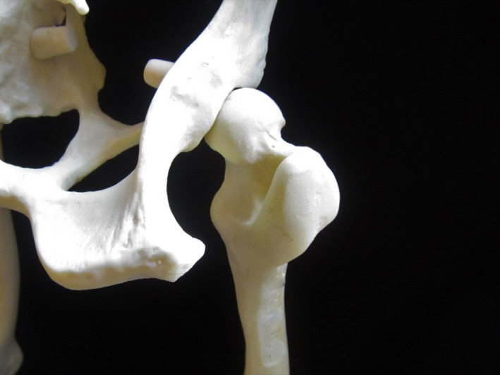
Landmarks for the sciatic nerve block on a model of a dog pelvis/femur. On the left is the ischial tuberosity and on the right is the greater trochanter of the femur. The injection is made 1/3 of the distance from the greater trochanter on a line that would connect the greater trochaner and the ischial tuberosity. Used for orthopedic and soft tissue surgeries of the pelvic limb.