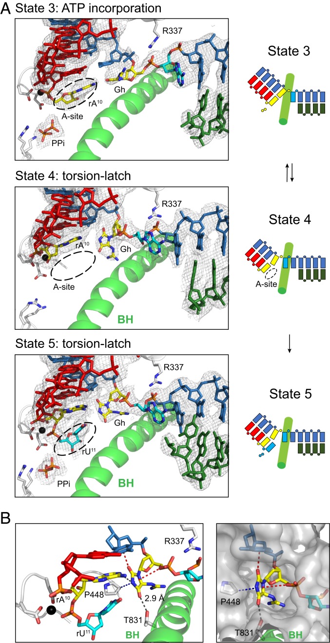
Fig. 8.
Unusual perpendicular Gh lesion reconfiguration impairs proper loading of the downstream undamaged 5′ template nucleobase during the extension step. (A) Structural analysis of consecutive translocation and extension of EC containing Gh lesion. (Left) Show canonical views of Pol II active sites in four states (states 3 to 6), with 2Fo-Fc electron density map of each scaffold contoured at 1.1 σ. In state 3, Gh lesion is located at canonical +1 template position and newly incorporated rA10 is located at the pretranslocated state. The pyrophosphate group is shown as PPi. In state 4, rA10 is translocated to −1 position, whereas the hydantoin ring of Gh rotates about 90°, thereby occupying both canonical −1 and +1 template positions. Crystal structures after UTP soaking were solved in two successive incorporations of UTP (SI Appendix, Fig. S5E) (states 5 and 6). In state 5, the rU11 is located at the pretranslocate state, whereas Gh occupies between the canonical −1 and +1 template positions. (Right) Schematic representation of extension which starts from state 3. (B) Detailed analysis of binding environment of rotated Gh base in state 5. Possible hydrogen bonding is indicated with black dashes. Lone pair–π and CH–π interactions are indicated with red and blue dashes, respectively. To indicate the binding pocket of rotated Gh, surface is shown in gray.

