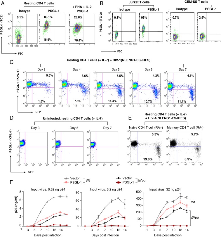
Fig. 1.
PSGL-1 is expressed on human CD4 T cells and exerts an anti-HIV activity. (A) Peripheral blood resting CD4 T cells were purified by negative selection, activated with PHA+IL-2, or left unstimulated. Cell surface PSGL-1 expression was analyzed by flow cytometry. (B) Jurkat and CEM-SS cells were similarly stained for surface PSGL-1. (C–E) Down-regulation of PSGL-1 by HIV-1 in primary CD4 T cells. Primary resting CD4 T cells were infected with NLENG1-ES-IRES, a GFP reporter virus. Following infection, cells were washed and cultured in complete medium plus IL-7 (2 ng/mL) to permit low-level viral replication. Surface PSGL-1 expression was analyzed at the indicated days. (C) Percentages of the GFP+ or GFP− cells with low or high PSGL-1 staining in each panel. PSGL-1 down-regulation was observed only in the HIV-infected (GFP+) cell population. (D) For controls, uninfected cells were similarly cultured in IL-7, and surface PSGL-1 expression was analyzed at the indicated days. Culturing of resting CD4 T cells in IL-7 did not lead to PSGL-1 down-regulation. (E) Cells were also stained for surface PSGL-1 expression on CD45RA+ (naïve) and CD45RA− (memory) CD4 T cells on day 7 and analyzed by flow cytometry. (F) HeLa JC.53 cells were stably transfected with PSGL-1 or empty vector DNA and drug-selected to obtain stably transfected cells. These cells were then infected with three different inputs of HIV-1(NL4-3)WT or HIV-1(∆Vpu). Viral replication was quantified by p24 release.

