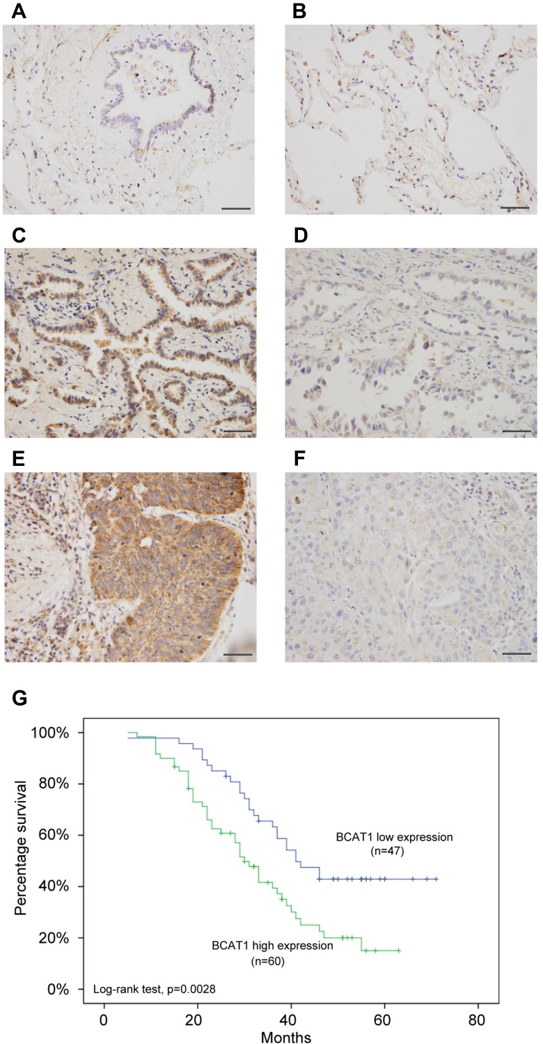
Figure 1.
BCAT1 expression is increased in lung cancer tissues. (A) Negative/weak immunostaining of BCAT1 in normal bronchial epithelial tissue. (B) Weak BCAT1 immunostaining in normal alveolar epithelial tissue. (C) Positive cytoplasmic BCAT1 expression in a case of lung adenocarcinoma. (D) Weak cytoplasmic BCAT1 immunostaining in a case of lung adenocarcinoma. (E) Strong cytoplasmic BCAT1 expression in a case of lung squamous cell carcinoma. (400x; bars indicate 50µm). (F) Negative/weak cytoplasmic BCAT1 expression in a case of lung squamous cell carcinoma. (G) Overall survival curve of lung cancer patients with high and low BCAT1 expression (Log-rank test, p=0.0028).

