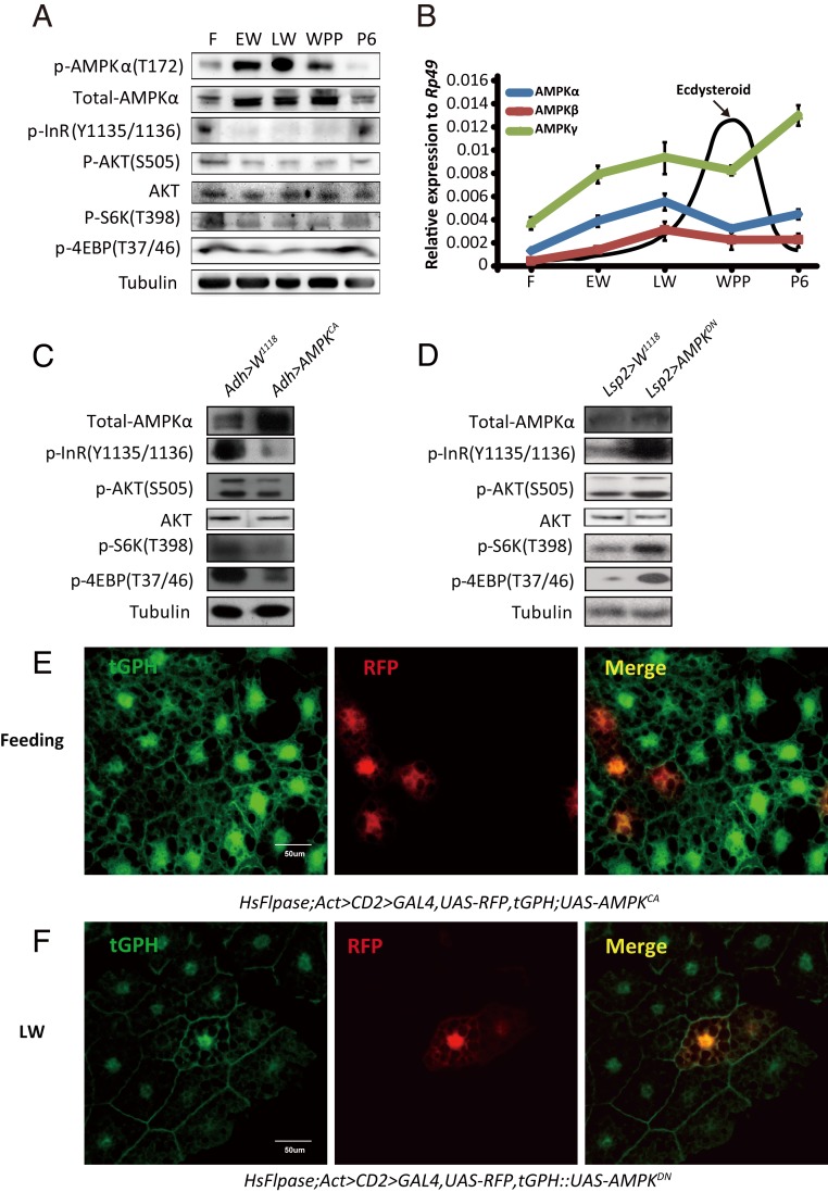
Fig. 2.
AMPK inhibits IIS in Drosophila. (A) Developmental phosphorylation profile of AMPK, AKT, InR, S6K, and 4EBP in the Drosophila fat body at different stages (feeding, EW, LW, WPP, and 6 h after pupation). (B) Developmental profiles of mRNA levels of AMPKα (blue), AMPKβ (red), and AMPKγ (green) in the Drosophila fat body and ecdysteroid titers (72, 73) at different stages. Fold-changes shown are relative to Rp49. (C) Phosphorylation levels of InR, AKT, S6K, and 4EBP were decreased in Adh-Gal4 > UAS-AMPKCA. Adh-Gal4 drives fat body-specific Gal4 expression. (D) Phosphorylation levels of InR, AKT, S6K, and 4EBP were increased in Lsp2-Gal4 > UAS-AMPKDN. Lsp2-Gal4 also drives fat body-specific Gal4 expression. (E) The Flp-out experiment revealing that the activity of PI3K is inhibited in red-positive clones in HsFlpase; Act > CD2 > Gal4, UAS-RFP, tGPH; UAS-AMPKCA at the feeding stage. RFP (red), tGPH (green). (F) The Flp-out experiment revealing that the activity of PI3K is increased in red-positive clones in HsFlpase; Act > CD2 > Gal4, UAS-RFP, tGPH::UAS-AMPKDN at the LW stage. RFP (red), tGPH (green).

