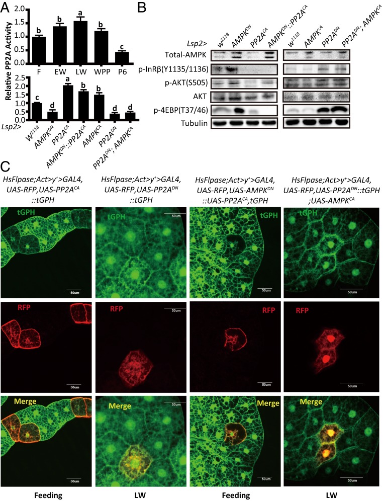
Fig. 3.
AMPK acts through PP2A to inhibit IIS in Drosophila fat body. (A, Upper) Developmental profile of the relative PP2A activity in the Drosophila fat body at different stages (feeding, EW, LW, WPP, and 6 h after pupation). Fold-changes shown are relative to the feeding stage; (Lower) Relative PP2A activities in the fat body of different Drosophila genotypes (W1118, Lsp2-Gal4 > UAS-AMPKDN, Lsp2-Gal4 > UAS-PP2ACA, Lsp2-Gal4 > UAS-AMPKDN::UAS-PP2ACA, Lsp2-Gal4 > UAS-AMPKCA, Lsp2-Gal4 > UAS-PP2ADN, and Lsp2-Gal4 > UAS-PP2ADN; UAS-AMPKCA). Fold-changes shown are relative to the W1118. Statistical significance between samples was evaluated using ANOVA: Bars labeled with different lowercase letters are significantly different (P < 0.05). (B) Western blot analysis results in the fat body of different Drosophila genotypes as in A, Lower. (C) A Flp-out experiment revealing that the activity of PI3K is decreased in red-positive clones in HsFlpase; Act > y+ > Gal4, UAS-RFP, tGPH::UAS-PP2ACA and HsFlpase; Act > y+ > Gal4, UAS-RFP, UAS-AMPKDN::UAS-PP2ACA, tGPH at the feeding stage, while the activity of PI3K is increased in HsFlpase; Act > y+ > Gal4, UAS-RFP, UAS-PP2ADN, and HsFlpase; Act > y+ > Gal4, UAS-RFP, UAS-PP2ADN::tGPH; UAS-AMPKCA at the LW stage. RFP (red), tGPH (green).

