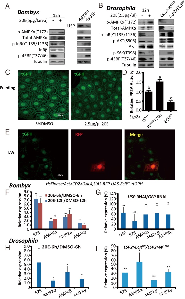
Fig. 4.
20E activates the AMPK-PP2A axis and inhibits IIS in insect fat body. (A) p-AMPKα, total-AMPKα, p-InR, and p-4EBP levels were measured after the 20E treatment (5 μg per larva) or USP RNAi in Bombyx. (A, Right) Anti-USP was used. (B) The levels of p-AMPKα, total-AMPKα, p-InR, p-AKT, p-S6K, and p-4EBP were measured in 20E-fed (2.5 μg/μL) or Lsp2 > EcRDN Drosophila. (C) tGPH membrane localization in the fat body of Drosophila following different treatments (5% DMSO and 2.5 μg/μL 20E). tGPH (green). (D) Relative PP2A activity in 20E-fed (2.5 μg/μL) or Lsp2 > EcRDN Drosophila. Fold-changes shown are relative to the W1118. Statistical significance between samples was evaluated using ANOVA: Bars labeled with different lowercase letters are significantly different (P < 0.05). (E) Flp-out experiment revealing that the activity of PI3K is increased in red-positive clones in HsFlpase; Act > CD2 > Gal4, UAS-RFP, tGPH::UAS-EcRDN at the LW stage. RFP (red), tGPH (green). (F and G) mRNA levels of AMPKα, AMPKβ, and AMPKγ in the fat body of Bombyx following 20E treatment (6 h and 12 h) or USP RNAi. Fold-changes shown are relative to DMSO treatment or GFP RNAi, respectively. (H and I) mRNA levels of AMPKα, AMPKβ, and AMPKγ in the fat body of Drosophila 6 h after the 20E treatment or Lsp2 > EcRDN. Fold-changes shown are relative to DMSO treatment or Lsp2 > W1118, respectively. Statistical significance between samples was evaluated using Student’s t test (*P < 0.05, **P < 0.01).

