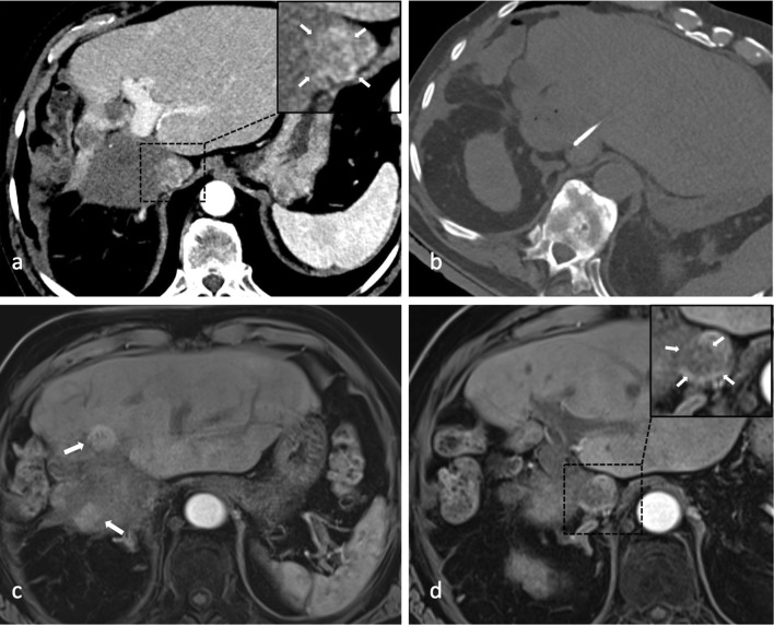
Fig. 3.
A Arterial phase CT showing contrast enhancing tumor thrombus (magnified image in the right upper corner with white arrows) in January 2018. B Axial non-enhanced control CT depicting needle placement for ablation of residual tumor thrombus in inferior caval vein (March 2018); last intervention up until now. C, D MRI scan of last follow-up in June 2019 with two recurrent nodules adjacent to necrosis zone (white arrows in c) and partially devascularized residual tumor thrombus in inferior caval vein (magnified image in the right upper corner with white arrows in D)

