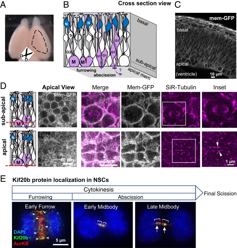
Fig. 1.
Imaging cytokinesis in NSCs of developing cortex. (A) Embryonic mouse brain with dashed outline showing area of cortex dissected for cortical slab preparation. (B) Schematic of NSCs forming the pseudostratified epithelium of the developing cerebral cortex. NSCs undergo interkinetic nuclear migration. Their nuclei move basally for S phase and apically for mitosis (M). Mitosis, furrowing, and abscission occur at the apical membrane. Abscission completes during G1 phase. N, postmitotic neurons. (C) Cross-section image of E12.5 mouse cortex expressing membrane-GFP shows the dense packing of NSCs in the epithelium. Round cells in mitosis can be seen close to the apical surface. (D) Schematics and images of cortical slabs labeled with membrane-GFP (green fluorescent protein) and SiR-Tubulin, viewing the apical membrane en face, at two different imaging planes (red dashed lines). Upper shows the subapical plane where the rounded mitotic cells with larger cell diameters and mitotic spindles are located. Lower shows the apical plane where apical endfeet and cell junctions are located and where the midbody forms and abscission occurs. Arrowheads point to the central bulges of two different midbodies. (Scale bars in D also apply to panels directly above.) (E) Endogenous Kif20b immunostaining in dissociated fixed embryonic cortical mouse NSCs shows changes in Kif20b localization (green) at progressive stages of cytokinesis. A cell in the early furrowing stage shows many Kif20b puncta (arrowheads) on the central spindle, which is labeled by AuroraB kinase (red). Chromatin is labeled by DAPI (4′,6-diamidino-2-phenylindole) (blue). A cell at early midbody stage of abscission shows Kif20b localized on the entire AuroraB-positive midbody flanks (brackets), but at late midbody stage, Kif20b becomes enriched on the outer flanks (arrows) near the constriction sites. Furrowing starts during anaphase, and abscission progresses during telophase and completes with final scission in G1. Kif20b staining is undetectable in Kif20b mutant NSCs (27). (Scale bar in E applies to all three images.) Image credit: Michael Fleming (University of Virginia School of Medicine, Charlottesville, VA).

