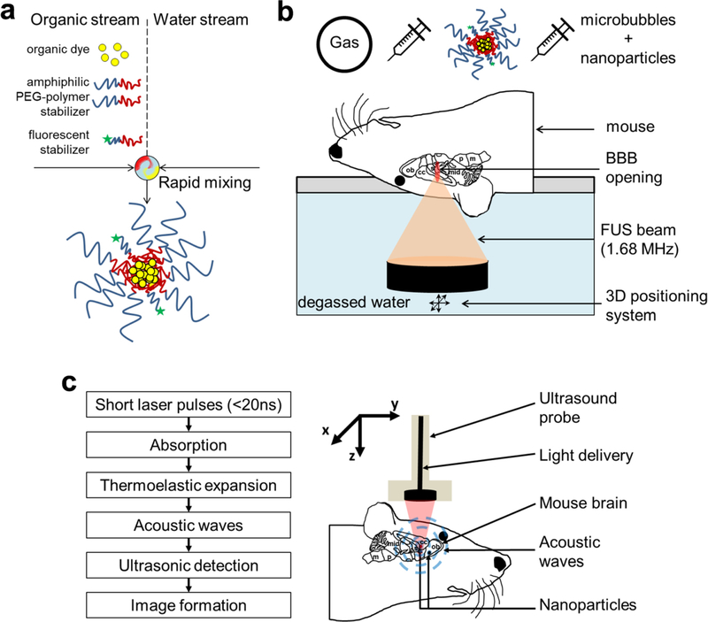
Fig. 1.
FNP processes, FUS combined with nanoparticle and schematic illustration of PAI in brain tissue. a FNP assembly of photoacoustic nanoparticles. b FUS system combined with circulating microbubbles allows BBB opening. c PAI consists of short laser pulses absorbed locally in tissue resulting in an increase in temperature leading to a thermoelastic expansion that creates acoustic waves detectable by US. Blood and NPs are strong absorbers in NIR laser wavelengths (680 nm to 970 nm) used; therefore, the vasculature or NPs within the tissue is observed. ob: olfactory bulb; cc: cerebral cortex; s: septum; mid: midbrain; p: pons and m: medulla.

