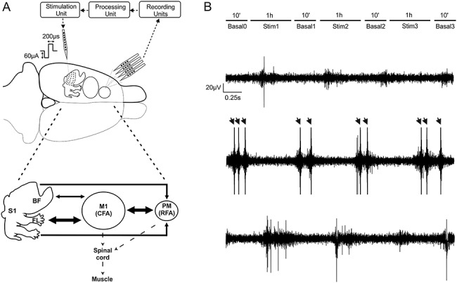
Figure 1.

Recording and stimulation experimental paradigm. (A) Extracellular recordings were obtained through a four-shank, 16-contact microelectrode probe in anesthetized rats. Signals were acquired (“Recording Unit”) and processed to detect single-unit activity (“Processing Unit”) selected by the user and employed as reference neuron to trigger a stimulus pulse to a channel on the single-shank microelectrode (“Stimulation Unit”). A summary diagram showing the main cortico-cortical anatomical connections between regions is shown in Fig. 1A, bottom. Arrow thickness corresponds to the amount of labeling (medium or high) suggested by Zakiewicz (Zakiewicz et al. 2014). (B) Recording sessions (in RFA) consisted of three 1-hour intermittent periods of stimulation with either ADS or RS to either S1BF or S1FL, each separated by 10-min periods of no stimulation. Example traces in a representative experiment of extracellular recording during the first basal period (Basal0, Top), during the first stimulation session (Stim1, Mid), and during the second basal period, after the stimulation (Basal1, Bottom). Arrows represent the electrical stimulation artifacts.
