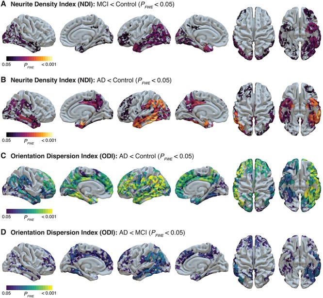
Figure 2.

Decreased cortical gray matter NODDI metrics in MCI and AD dementia groups from whole-brain GBBS analysis. NDI in cortical gray matter is significantly decreased in both MCI (A) and AD dementia (B). ODI in gray matter is unchanged in MCI but significantly decreased in AD dementia relative to both the control group (C) and the MCI group (D). GBSS statistical maps were projected onto surfaces in Surf Ice and indicate areas with significant (FWE-corrected P < 0.05) differences. Representative axial slices of significant GBSS results can be found in Supplementary Figure 1.
