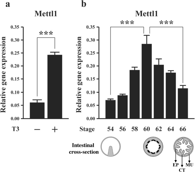
Fig. 1.
Expression of Xenopus tropicalis Mettl1 increases during T3-induced and natural metamorphosis. a The expression of Mettl1 was analyzed during T3-induced metamorphosis. Stage 54 tadpoles were treated with 10 nM T3 for 2 days and total RNA was isolated from the intestine for RT-PCR analysis. b During natural metamorphosis, Mettl1 expression gradually increased from premetamorphic period to peak at the metamorphic climax. Total RNA was isolated from the intestine of tadpoles at indicated stages for RT-PCR analysis. Shown below the expression data are schematic diagrams of the intestine at different stages. In premetamorphic tadpoles at stage 54, the intestine is a simple structure with a single epithelial fold, the typhlosole, and thin layers of connective tissue and muscles. At the metamorphic climax around stage 61, the larval epithelial cells begin to undergo apoptosis, as indicated by the open circles. Concurrently, the proliferating adult stem cells are formed de novo via dedifferentiation of some larval epithelial cells, as indicated by black dots. By the end of metamorphosis at stage 66, the newly developed adult epithelium (EP) has multiple folds, surrounded by thick layers of connective tissue (CT) and muscles (MU). L: intestinal lumen. All data represent mean ± S.E.M. Significance value was ***P ≤ 0.005

