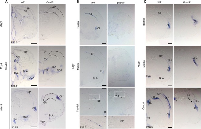
Figure 4.

SP defects are more severe in the rostral than in the caudal part of the cortex of Dmrt5−/− embryos. (A–C) ISH for Pls3, Pcp4, and Nurr1 in coronal sections from WT and Dmrt5−/− embryos at E15.5 (A) and for Ctgf and Nurr1 at E18.5 (B and C). Expression of the SP markers Pcp4, Pls3, and Nurr1 is strongly reduced in the neocortex and Hpc at E15.5 in Dmrt5−/−. At E18.5, Ctgf and Nurr1 expressions were however detected in the SP within the caudal part of the cortex (black arrowheads). Higher magnification of the boxed area is shown for Ctgf staining in caudal sections. dLn: deep layers neurons; EN: endopiriform nucleus; VZ: ventricular zone. Scale bar represents 500 μm for all low power images in A–C and 100 μm for lower panels in B.
