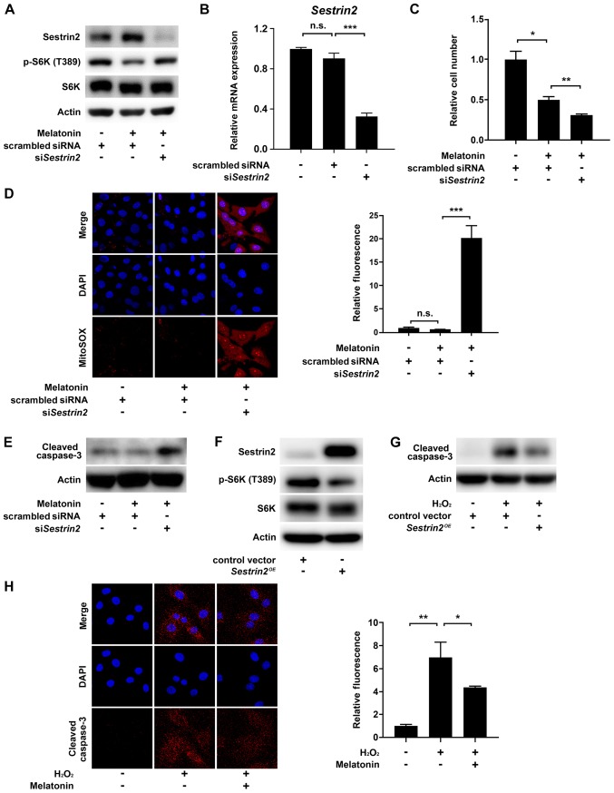
Figure 4.
Effects of si-Sestrin2 on ROS generation and apoptosis in melatonin-treated VSMCs. Primary rat VSMCs transfected with scrambled siRNA or siSestrin2 for 24 h were serum-starved for 24 h, and then incubated with 10% FBS with or without 2 mM melatonin. Levels of (A) phosphorylated S6K (T389) and (B) Sesn2 mRNA in VSMCs. (C) Relative VSMC number. Data are presented as the mean ± SEM. (D) Representative fluorescence micrographs showing intracellular superoxide production in VSMCs. Bar graph shows quantitation of MitoSOX fluorescence intensity (red) with nuclear counterstaining with DAPI (blue), expressed as the mean ± SEM. (E) Level of cleaved caspase 3 protein in VSMCs. (F and G) Primary rat VSMCs transfected with the Sestrin2-expression vector for 24 h were serum-starved for 24 h, and then incubated with or without 1 mM H2O2 for 6 h. (F) Phosphorylated S6K (T389) and (G) cleaved caspase 3 protein levels in VSMCs. (H) Primary rat VSMCs were pretreated with 2 mM melatonin for 24 h and then treated with or without 1 mM H2O2 for 6 h. Immunofluorescence images of cleaved caspase-3 (red) with nuclear counterstaining with DAPI (blue). Bar graphs show quantitation of cleaved caspase-3 immunofluorescence intensity, expressed as the mean ± SEM. Magnification, x100. *P<0.05; **P<0.01; ***P<0.001. n.s., not significant; VSMC, vascular smooth muscle cell; si/siRNA, small interfering RNA; ROS, reactive oxygen species.

