Figure 2.
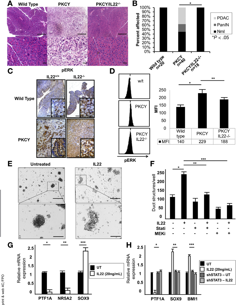
IL22 promotes PDAC initiation through Stat3-mediated induction of ADM. (A) Representative H&E of murine pancreata from WT, PKCY, and PKCY/IL22−/− mice (top row scale bars, 400 μm; bottom row scale bars, 40 mm). (B) Incidence of PanlNs and invasive cancer in WT, PKCY, and PkCy/IL22−/− mice (*P < .05). (C) IHC staining showed lower intensity of pERK in PKCY/IL22−/− compared with PKCY mice (n = 5) (scale bars, 40 μm). (D) Intensity of pERK before tumor formation by flow cytometric analysis of EpCAM+ cells isolated from the pancreas (representative fluorescence-activated cell sorting data shown, n = 5; *P < .05, **P < .05). (E, F) IL22 (20 ng/mL) treatment of acinar cultures for 48 hours induced acinar-to-ductal metaplasia, which was abrogated by inhibition of JAK/Stat (ruxolitinib, 2.5 mmol/L) and MEK (trametinib, 0.1 mmol/L) (*P < .05, **P < .05, ***P < .05) (scale bars, 400 μm). (G, H) qRT-PCR showed that 24 hours of IL22 treatment enhanced the expression of the ductal marker SOX9, which was dependent on Stat3 (*P < .05, **P < .05, ***P < .05). MFI, mean fluorescence intensity; Nml, normal; sh, short hairpin; UT.

