
Figure 4.
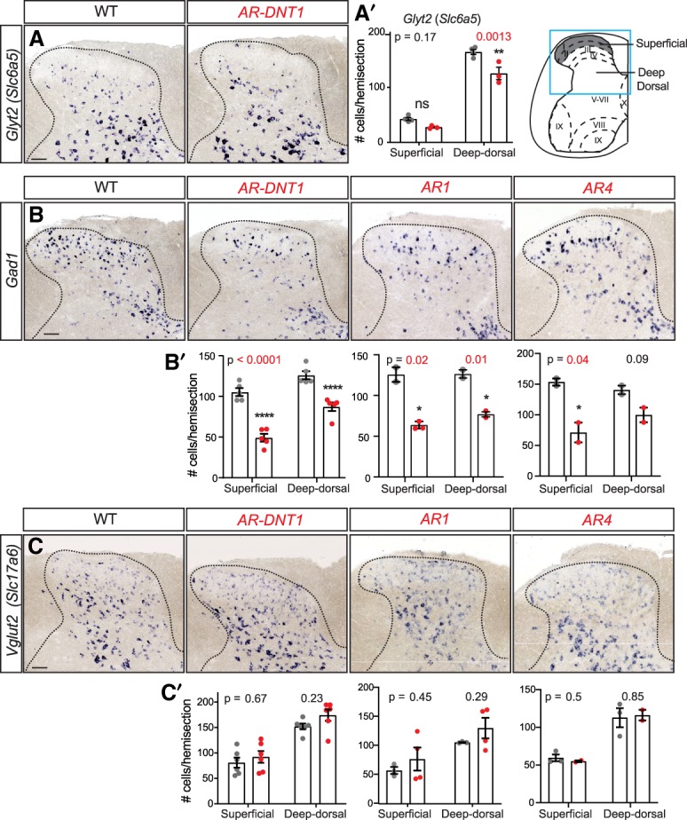
Reduced PTF1A levels affect specification of Gad1 and Glyt2 (Slc6a5) inhibitory neurons. (A–C) ISH shows a decrease in Glyt2 (A) and Gad1 (B), but no significant change in VGlut2 (Slc17a6) (C) neurons in Ptf1a enhancer mutants with the scratch phenotype (AR-DNT1, AR1, and AR4) at P30 compared with WT. (A′–C′) Quantification reports the number of marker+ cells per hemisection from superficial (gray in diagram) or deep dorsal spinal cord regions for the markers shown in A–C, respectively. Each data point represents a biological replicate (N), error bars indicate SEM. Student's t-test was used to determine significant differences relative to WT, P-values are as indicated. (*) P < 0.05; (**) P < 0.01; (****) P < 0.0001. Scale bar, 100 µm. See also Supplemental Fig. S4.