Figure 2.
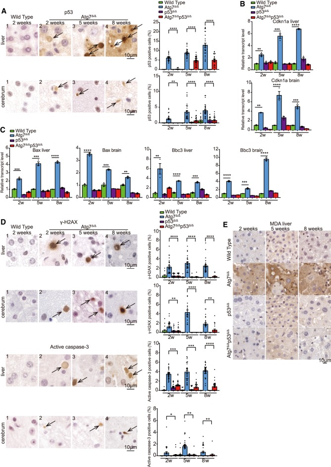
Autophagy is required to protect liver and brain from p53 accumulation, DNA damage response activation, and apoptosis. (A) Representative liver and cerebrum IHC staining of p53 and quantification at the indicated times from wild-type and Atg7Δ/Δ mice. Black arrows indicate p53-positive cells. (2w) 2-wk time point; (5w) 5-wk time point; (8w) 8-wk time point. (B,C) Quantitative real-time PCR of Cdkn1a, Bax, and Bbc3 for liver and brain tissues from wild-type, Atg7Δ/Δ, p53Δ/Δ, and Atg7Δ/Δp53Δ/Δ mice at the indicated times. (**) P < 0.01; (***) P < 0.001; (****) P < 0.0001 (unpaired t-test). (D) Representative liver and cerebrum IHC staining for γ-H2AX and active caspase-3 with quantification at the indicated times from wild-type and Atg7Δ/Δ mice. Black arrows indicate γ-H2AX or active caspase-3-positive cells. (2w) 2 wk; (5w) 5 wk; (8w) 8 wk. (*) P < 0.05; (**) P < 0.01; (***) P < 0.001 (****) P < 0.0001(unpaired t-test). (E) Representative liver IHC staining for MDA at the indicated times from wild-type, Atg7Δ/Δ, p53Δ/Δ, and Atg7Δ/Δp53Δ/Δ mice. See also Supplemental Figure S2.

