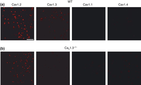
Figure 1.

Cav1 channel subtypes expressed in mouse chromaffin cells. Immunocytochemical characterization of Cav1 channel subtypes. (a–b) Confocal images of isolated mouse chromaffin cells from WT (a) or Cav1.3−/− mice (b) labeled with antibodies against Cav1.1, Cav1.2, Cav1.3 and Cav1.4 channels (dilution 1 : 200) and the corresponding secondary antibody (dilution 1 : 200) Alexa Fluor excited at a wavelength of 594 nm (dilution 1 : 200). Experiments were performed on four paired cultures of WT and Cav1.3−/− cells. Calibration bar: 75 microns.
