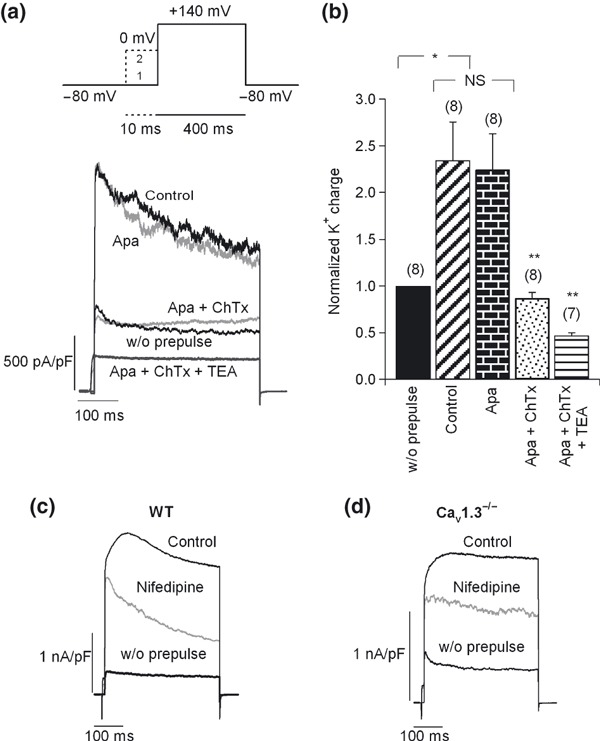
Figure 7.

Coupling of Cav1 channel subtypes to BK channels. (a) Upper section: double‐pulse protocol used to recruit BK channels. This included a 400 ms test pulse (V t) to 140 mV or above that potential (trace 1), followed by a 10‐ms pre‐pulse applied at 0 mV before V t (trace 2). The Ca2+ dependent K+ currents activated using this protocol were BK channels. Lower section: original K+ current traces recorded using the above protocol under control conditions and after perfusion with different K+ channel blockers, added sequentially and cumulatively: first, 200 nM apamin, 100 nM charibdotoxin (ChTx), and finally 45 mM TEA. Pulses were applied every 2 min. Numbers of cells are indicated in parentheses. (b) The K+ charge density was averaged and normalized for each condition with respect to the current in the absence of a pre‐pulse. (c–d) Effects of 3 μM nifedipine on BK channel currents in WT (c) and Cav1.3−/− cells (d). Number of cells: 14 WT cells, 11 Cav1.3−/− cells. Data were obtained in four paired cultures of WT and Cav1.3−/− cells, using two mice of each strain.
