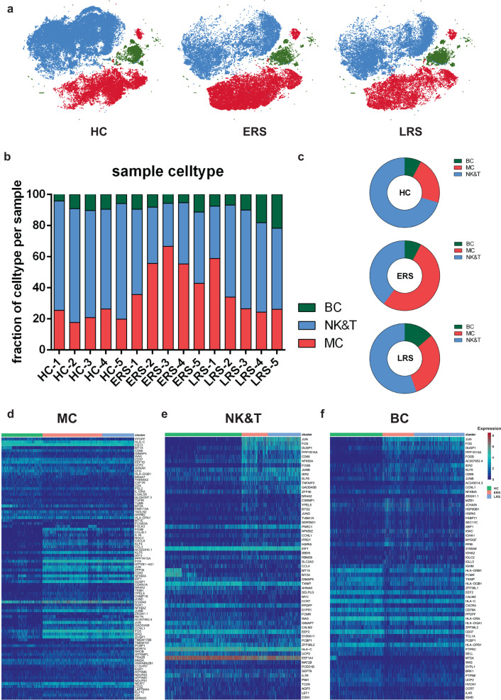
Fig. 2. An overview of NK and T, B, and myeloid cells in the blood of convalescent patients with COVID-19.
a The t-SNE plot shows a comparison of the clustering distribution across HCs as well as early recovery stage (ERS) and late recovery stage (LRS) patients with COVID-19. b The bar plot shows the relative contributions of myeloid, NK and T, and B cells by individual samples, including five HCs, five ERS patients, and five LRS patients. c The pie chart shows the percentages of myeloid, NK and T, and B cells across HCs as well as ERS and LRS patients with COVID-19. d The heatmap shows the DEGs of myeloid cells among the HCs and the ERS and LRS COVID-19 patients. e The heatmap shows the DEGs of NK and T cells among the HCs and the ERS and LRS COVID-19 patients. f The heatmap shows the DEGs of B cells among the HCs and the ERS and LRS COVID-19 patients.

