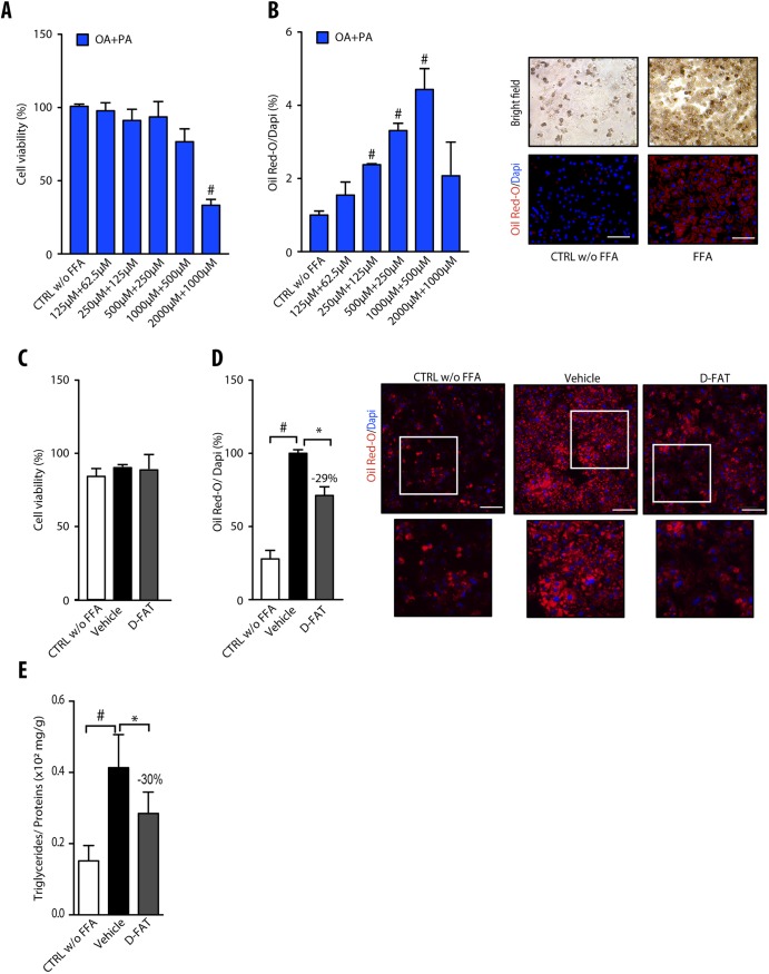
Fig. 1.
Steatosis induction and defatting response to D-FAT in PHH. (A,B) Normal human hepatocytes in primary culture were incubated with or without different concentrations of the free fatty acid (FFA) mixture oleic acid (OA) and palmitic acid (PA) (2:1) for 48 h, and examined for (A) cell viability, assessed by MTT assay, and (B) lipid droplet content, assessed by Oil Red O staining. In B, left panel shows quantification of Oil Red O staining, normalized to the number of DAPI-stained nuclei; right panel shows representative images of controls (CTRL) and FFA-loaded PHH (OA:PA, 500:250 μmol/l). (C-E) Normal human hepatocytes in primary culture were incubated with or without FFA mixture (OA:PA, 500:250 μmol/l) for 48 h, and thereafter FFA-loaded PHHs were treated with the D-FAT cocktail or the vehicle for 24 h, and then subjected to (C) cell viability, assessed via the MTT assay; or assessed for (D) lipid droplet content, by Oil Red O staining; or (E) intracellular triglyceride (TG) content normalized for cell protein. In D, left panel shows quantification of Oil Red O staining; right panel shows representative images of controls and FFA-loaded PHH at baseline, and after vehicle or DFAT treatment (lower panels show magnification of boxed areas in upper panels). Means±s.e.m. of six cell preparations are shown relative to controls in A and B and to vehicle in D and E. In all panels, #P<0.05 versus control, *P<0.05 versus vehicle (one-way ANOVA). Scale bars: 50 μm.

