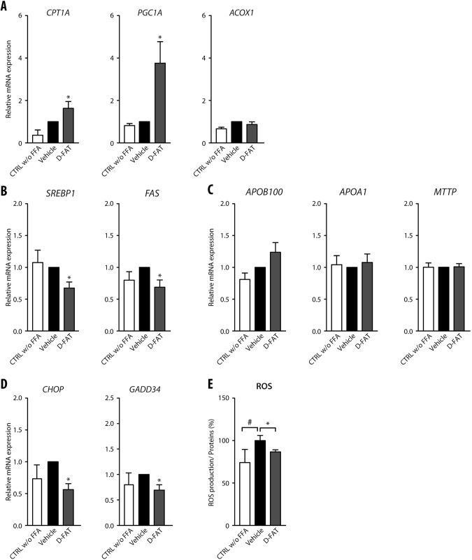
Fig. 2.
Pathways altered along with defatting in response to D-FAT in PHH. (A-E) Normal human hepatocytes in primary culture were incubated with or without FFA mixture (OA:PA, 500:250 µmol/l) for 48 h, and thereafter FFA-loaded PHHs were treated with the D-FAT cocktail or the vehicle for 24 h. PHHs in all conditions were subjected to RT-qPCR analyses of genes involved in fatty acid β-oxidation (A) (CPT1A, PGC1A, ACOX1); lipogenesis (B) (SREBP1, FAS); lipid export (C) (ApoB100, ApoA1, MTTP); and ER stress (D) (CHOP, GADD34). (E) Measurement of intracellular ROS by spectrophotometric quantification of H2DCFDA. Data are mean±s.e.m. of six cell preparations, shown relative to vehicle. #P<0.05 versus control, *P<0.05 versus vehicle (one-way ANOVA).

