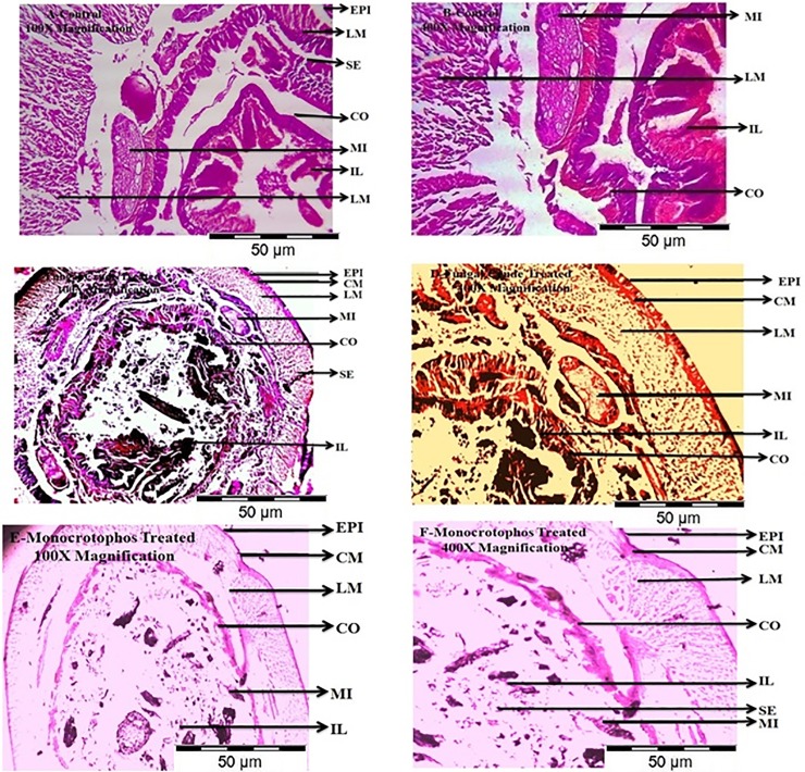
Fig 8. Mid gut cross section of the earthworm, E. eugeniae exposed to M.anisopliae secondary metabolites for 24hr post treatment with 500μg/ml concentration.
E. eugeniae cross section was magnified at 100X and 400X under light microscope. A and B is control; C and D is Monocrotophos 500μg/ml treated; E and F is to M.anisopliae extract treated. Among the control and treated few histopathological changes was observed in intestinal lumen and intestine was totally damaged compare with control (EPI-epidermis; CM-circular muscle; LM-longitudinal muscle; SE-setae; CO-coelom; MI-mitochondrion; IL-intestinal lumen).

