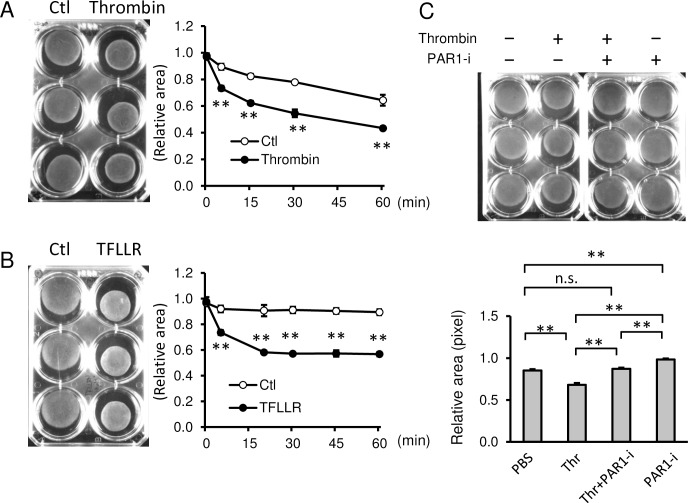
Fig 2.
Thrombin increased contraction of primary human myometrial cells through PAR1. (A and B) (Left images) Representative images of collagen lattice assay of human myometrial cells at 30 min treated with PBS (Ctl) and thrombin (A) or PAR1 activating peptide, TFLLR (B). (Right graphs) Quantification of myometrial contractions in collagen lattice assays (n = 3). **, p < 0.01 at each time point. (C) Collagen lattice assay of myometrial cells at 30 min with 2 U/mL of thrombin (Thr) pretreated with or without 100 nM PAR1 selective inhibitor (SCH79797, PAR1-i) for 1 h (n = 3). Representative image (upper panel) and quantification of gel areas (lower graph). The experiments were repeated three times. *, p < 0.05, and **, p < 0.01.

