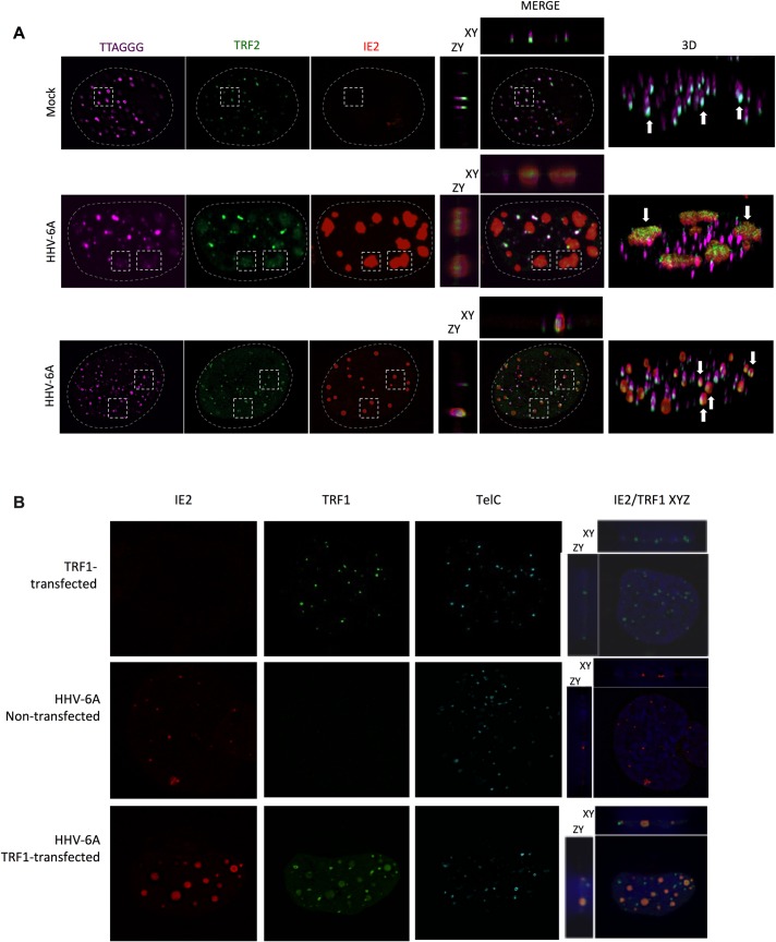
Fig 5. Colocalization of shelterin complex proteins and HHV-6A IE2 protein at VRC and cellular telomeres.
A) U2OS cells were mock-infected or infected with HHV-6A for 48h after which cells were processed for IF-FISH. Telomeres were labeled in magenta, TRF2 in green and IE2 in red. The panels in the middle row show images of cells with IE2 patches overlapping with large, diffuse TRF2 and telomeric staining (rectangles). The panels in the third row represent infected cells with punctate IE2 pattern colocalizing with TRF2 and telomeres (dashed squares). The colocalization of IE2, TRF2 and telomeres are shown in both 2D and 3D images. B) Uninfected and HHV-6A-infected U2OS cells were transfected with an empty vector, a myc-tagged-TRF1 expression vector. Forty-eight hours later cells were processed for IF-FISH. TRF1 was labeled in green and IE2 in red. Nuclei were stained with DAPI. Images on the far right show 2D colocalization of TRF1 with IE2.

