Fig 6.
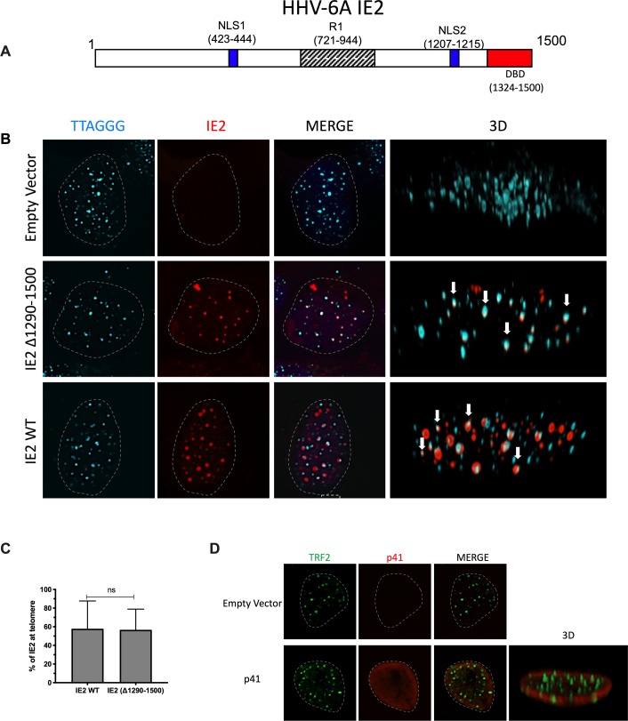
A) A stick diagram of the IE2 protein with various domains identified is presented. B) Colocalization of HHV-6A IE2 protein with telomeres in the absence of viral DNA. U2OS cells were transfected with an empty vector, with IE2 expression vector or with IE2 Δ1290–1500 expression vector. Forty-eight hours later cells were processed for dual color immunofluorescence. Telomeres were labeled in cyan and IE2 in red. Nuclei are outlined by dashed lines. Examples of IE2 colocalizing with telomeres are presented in a 3D view (white arrows). C) The graph represents the mean ± SD % of WT IE2 and Δ1290–1500 IE2 localizing with telomeres. D) Lack of colocalization between HHV-6A p41 and telomeres in uninfected cells. U2OS cells were transfected with an empty vector or with a p41 expression vector. Forty-eight hours later cells were processed for dual color immunofluorescence. TRF2 was labeled green, p41 in red and nuclei outlined by a dashed line.

