Fig. 5.
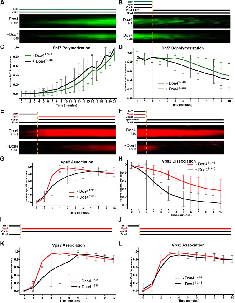
Doa4 inhibits Snf7 polymer association with Vps2:24 in vitro. (A) Experimental setup and kymographs from time-lapse fluorescence microscopy of representative Snf7-AlexaFluor-488 patches grown on supported lipid membranes with (+) or without (−) Doa41-348. (B) Kymographs of representative Snf7 patch disassembly. Snf7-AlexaFluor-488 was polymerized together with Vps2 and Vps24. At t=0, these were washed out, followed by the addition of ATP and Vps4. (C,D) Quantification of mean Snf7 fluorescence during polymerization (C) and depolymerization (D) of Snf7 in the presence (+) or absence (−) of Doa41-348, of at least 58 patches and 3 independent experiments from A and B, respectively. (E) Kymographs of Vps2 associating with representative Snf7 patches polymerized on supported lipid membranes. At t=0, Snf7 was washed out and Vps2-Atto-565 along with Vps24 was added. (F) Kymographs of Vps2 dissociation from representative Snf7 patches polymerized with Vps2-Atto-565 and Vps24 on supported lipid membranes. At t=0, these were washed out, followed by the addition of ATP and Vps4. (G,H) Quantification of mean Vps2 fluorescence during association (G) and dissociation of Vps2 (F) in the presence (+) or absence (−) of Doa41-348, of at least 56 patches and 3 independent experiments each from E and F, respectively. (I,J) Experimental setup for Vps2 associating with representative ESCRT-III patches. Snf7 was polymerized on supported lipid membranes. At t=0, Vps2-Atto-565 together with Vps24 was added. Doa41-348 was added either at t=0 (I) or during the initial Snf7 polymerization and washed out at t=0 (J). (K,L) Quantification of mean Vps2 fluorescence during Vps2 association in the presence (+) or absence (−) of Doa41-348 of at least 91 patches and 4 independent experiments each, from set-up described for I (K) and set-up described for J (L). Error bars in all graphs represent ±s.d.

