Fig. 1.
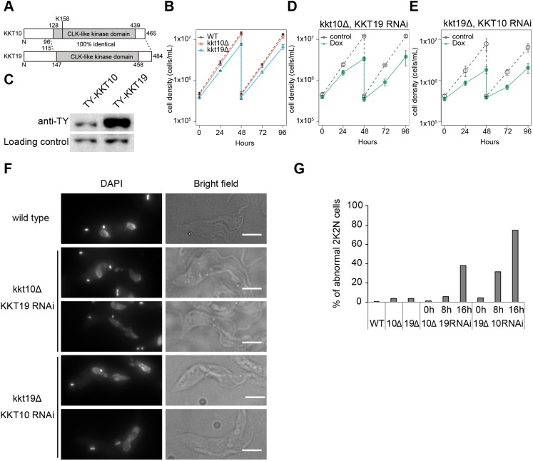
KKT10 and KKT19 are redundant. (A) Schematic representation of KKT10 and KKT19 proteins. (B) KKT10 (red) and KKT19 (blue) knockout cells are viable. Gray dashed line indicates a WT control. Results are mean±s.d. from three independent experiments. Similar results were obtained from at least two independent clones. (C) Protein levels of TY-YFP-tagged KKT10 and KKT19 were monitored by immunoblotting. Representative of three independent experiments is shown. PFR2 was used as a loading control. Uncropped images are shown in Fig. S1. (D,E) RNAi-mediated knockdown of (D) KKT19 in kkt10 deletion cells and (E) KKT10 in kkt19 deletion cells affects cell growth. Control is an uninduced cell culture. Results are mean±s.d. from three independent experiments. (F,G) KKT10/19 double depletion causes chromosome missegregation. (F) Examples of anaphase cells fixed at 16 h post induction of RNAi and stained with DAPI. Note that in WT cells, daughter nuclei appear as a smooth shape without significant lagging DNA in between them. In contrast, lagging chromosomes and/or abnormal nuclear signals were often observed upon depletion of KKT10/19. Maximum intensity projections are shown. Scale bars: 5 µm. (G) Quantification of the percentage of 2K2N cells with abnormal chromosome segregation (n≥74).

