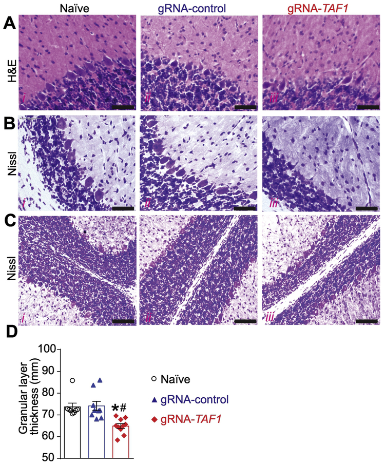
Figure 5. TAF1 gene editing results in morphologic changes in the cerebellum loss.
The morphology of the Purkinje cell layer was evaluated by Haemotoxylin and Eosin (A) and Nissl (B) Staining. Both H&E and Nissl staining showed abnormal architecture of the Purkinje cells as well as hypoplastic Purkinje cells. Scale bars: 500 μm. (C) The thickness of the Granular layer was evaluated by Nissl Staining. Nissl staining showed reduced granular layer thickness in gRNA-TAF1 group. Scale bars: 200 μm. (D) Summary of morphometric analysis of the Granular layer thickness. Data are shown as mean ± S.E.M., n = 9 fields per animal, 3 animals per experimental condition. *p<0.05 versus; naïve, #p<0.05 versus gRNA-control (ANOVA followed by Tukey’s test). The experiments were conducted in a blinded fashion.

