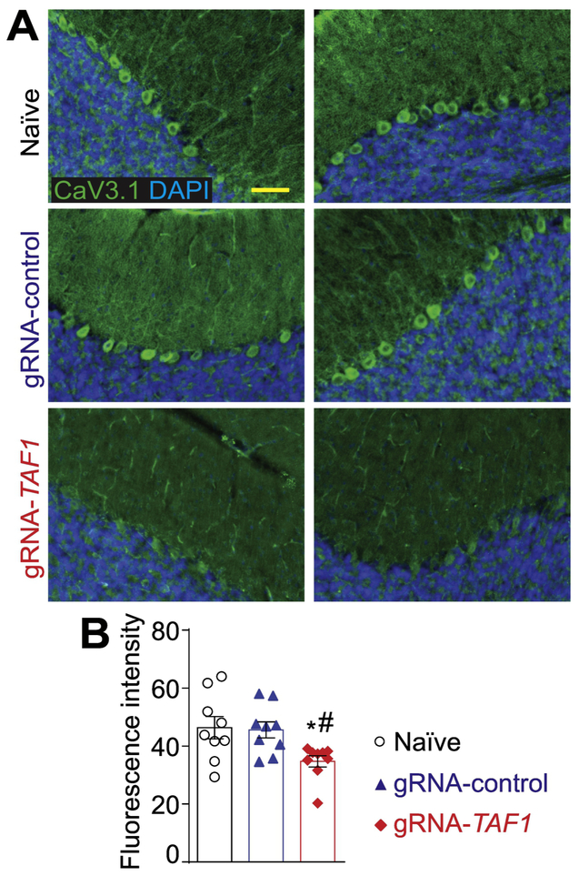
Figure 8. TAF1 gene editing leads to a downregulation of the CaV3.1 voltage-gated calcium channel.
(A) Representative photomicrographs of cerebellar slices from animals injected with (vehicle) or control or TAF1 gRNAs stained with an antibody against CaV3.1. Nuclei stained with DAPI. (B) Quantification of fluorescence intensity for the CaV3.1 immunohistochemistry. Scale bar = 200 μm. The experiments were performed in a blinded fashion. Data are shown as mean ± S.E.M. from 3 different fields from 3 animals per experimental condition (i.e. a total of nine fields per group). *p<0.05 versus; naïve, #p<0.05 versus gRNA-control (ANOVA followed by Tukey’s test). The experiments were conducted in an investigator blinded manner.

