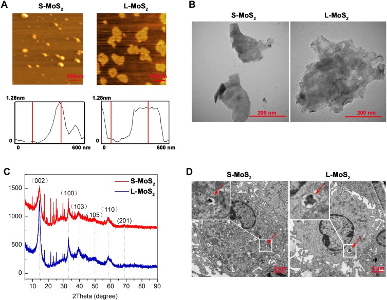
Figure 1.
Characterization of the few-layered MSNs and their uptake by DCs.
Notes: (A) AFM images of MSNs. (B) TEM images of MSNs. (C) The XRD pattern of MSNs. (D) DCs were incubated with MSNs (128 µg/mL) for 48 h and observed by TEM to examine the cellular uptake of MSNs. The red arrow indicates the internalized MSNs.
Abbreviations: S-MoS2, small MSNs; L-MoS2, large MSNs; AFM, atomic force microscopy; XRD, X-ray diffraction; TEM, transmission electron microscopy; MSNs, MoS2 nanosheets; DCs, dendritic cells.

