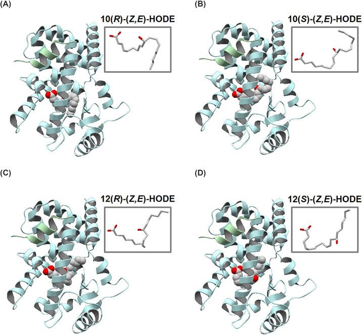
Figure 2. Overall structures of PPARγ complex with HODE isomers.
Docking poses of (A) 10(R)-(Z,E)-, (B) 10(S)-(Z,E)-, (C) 12(R)-(Z,E)-, and (D) 12(S)-(Z,E)-HODEs with the best S score (shown in Table 1), based on PPARγ protein from 2PGR. PPARγ and SRC1 are colored light blue and green, respectively, and HODEs are shown in space-fill model. The conformations of HODEs with the best S scores are shown in stick models at upper-right regions.

