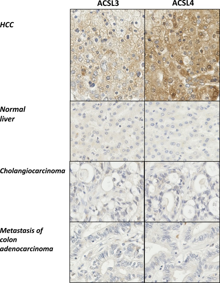
Figure 1. Immunohistochemistry reveals increased expression of both ACSL3 and ACSL4 in HCC.
Multiple liver tissues arrays were probed with antibodies specific for either ACSL3 or ACSL4. Representative examples (×20 maginfication) are shown for either ACSL3 or ACSL4 immunohistochemical staining of matched samples of HCC, normal liver, CCA and liver metastases.

