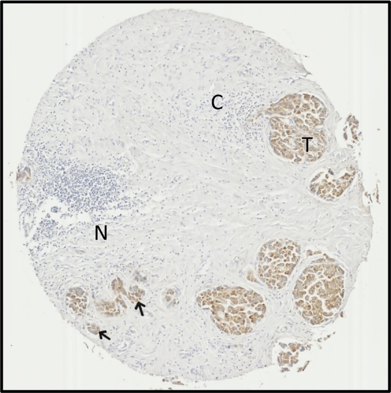
Figure 5. ACSL4 staining highlights tumour tissue amidst extensive cirrhosis and necrosis in samples.
Tissue microarray sample from a 60-year-old male with stage II HCC. ACSL4 staining is apparent in tumour regions (T) and small tumour tissue foci (arrows), but absent from regions of cirrhotic tissue (C) or necrosis (N). Images are ×5 magnified.

