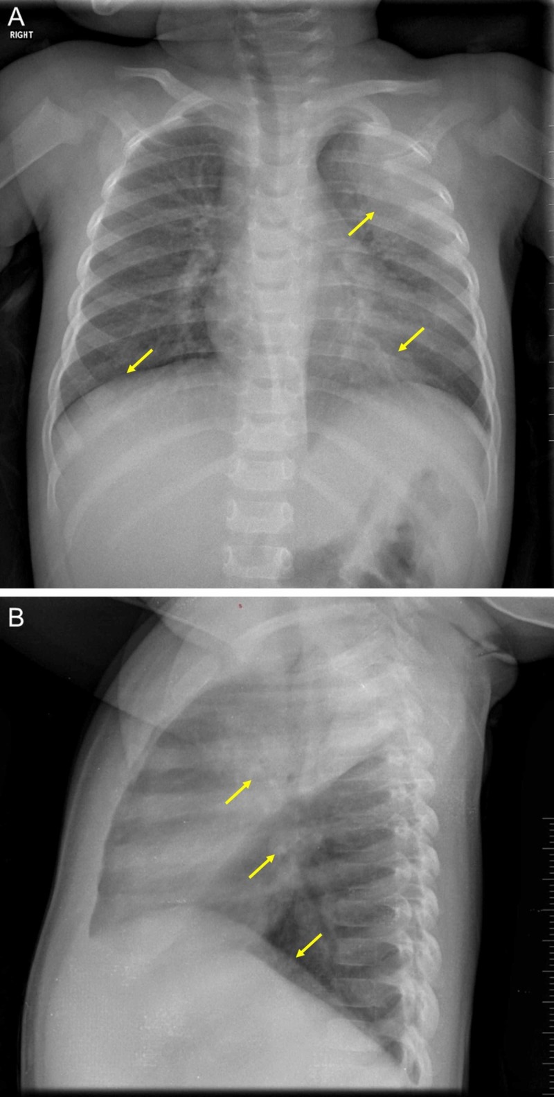
Figure 1. A chest radiograph.
Imaging of the infant’s chest shows large consolidation at the left apical lobe with bronchial infiltrates that are dominant at the left base, and asymmetrical lung bases (A). The heart size is normal, the rib cage is intact, and the diaphragmatic arches are in normal position (A). A discrete blunting at the left pleural sinus can be observed (B).

