
Figure 1. The anatomic topology of primary and recall T cell responses.
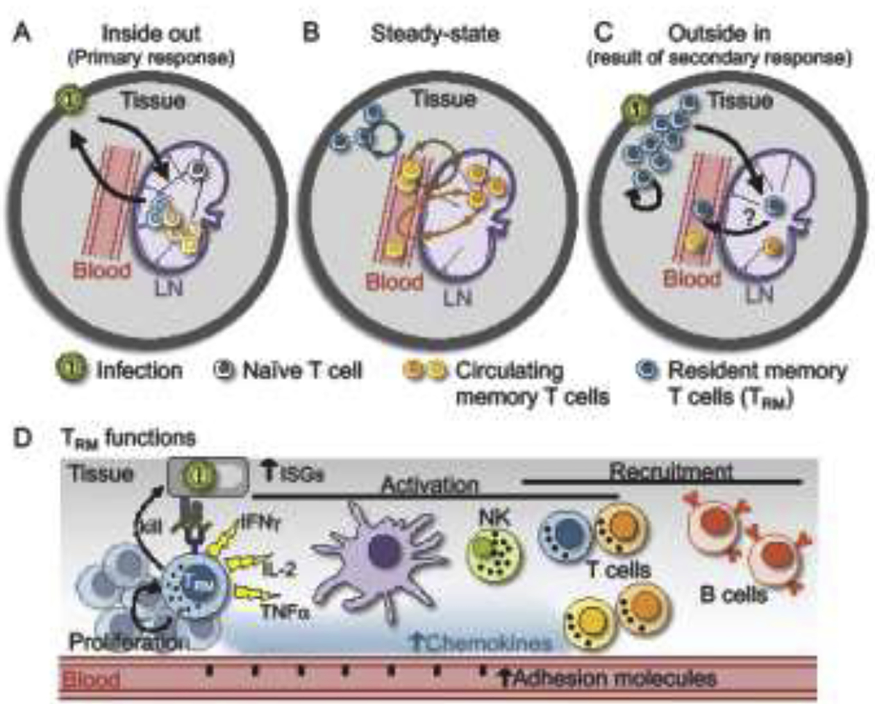
A) During a primary infection, antigen drains from peripheral tissues to draining lymph nodes (LN) which activates naïve T cells specific for that pathogen. Cells expand in lymphoid tissue and migrate out to peripheral tissues to control the infection (which could be referred to as an inside-out immune response). B) At steady-state, memory T cells patrol for reinfection and are heterogeneous, consisting of resident cells that are absent from circulation (TRM), cells that circulate through blood and lymph, and cells that recirculate though tissue using blood and lymph as conduits. C) Following secondary exposure to antigen, TRM can proliferate, give rise to an expanded local population of long-lived progeny, redistribute to draining LNs where they may remain resident, and possibly rejoin the circulation. TRM redistribution from the periphery to LNs and the circulating pool could be referred to as an outside-in immune response. Not pictured: Reactivation of LN circulating memory T cells (i.e., TCM) recapitulates an inside-out immune response. D) Upon sensing cognate antigen within tissue, TRM reactivate and alert the local immune system of a reinfection through chemokine and cytokine production. This leads to upregulation of interferon stimulated genes (ISGs), maturation of DCs, activation of T cells and NK cells, adhesion molecule upregulation and recruitment of CD8+ T cells and B cells. TRM also proliferate and upregulate cytotoxic molecules, likely contributing to killing of infected cells.